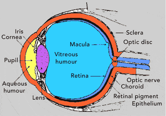
Before light can reach the rods and cones of the retina, it must pass through the cornea, aqueous humor, pupil, lens and vitreous humor. The first step in Physiology of Vision is retinal image formation and activation of Photoreceptors. The resulting nerve impulses are then passed to the visual areas of the cerebral cortex.
Retinal Image Formation:
This requires four basic processes,
- Refraction of light rays.
- Accumulation of the lens.
- Constriction of pupil.
- Convergence of eyes.
Light rays entering the eye from the air are refracted at the anterior surface of the cornea, posterior surface of the cornea, anterior surface of the lens and posterior surface of the lens. The degree of refraction that takes place at each surface is very precise and such that rays fall at the fovea centralis.
The lens of the eye is Biconvex. Furthermore, it has the unique ability to change the focusing power of the eye by becoming moderately curved at one moment and greatly curved the next. This change in curvature of lens is known as accommodation. In far vision, the ciliary muscle contracts pulling the ciliary process and choroid forward towards the lens. This causes shortening, thickening and bulging of the lens and thereby increasing the curvatures.
Constriction of pupil occurs in response to light reflex involving automonic nervous system and it is purely the function of smooth muscles of iris i.e. constrictor or circular muscles of iris.
Convergence in Physiology of Vision:
Human eyes are such that they focus on only one set of the object (single binocular vision). This type of vision is possible due to the phenomenon called Convergence. Convergence refers to the medial movement of the two eye-balls so that they are directed towards the object being viewed.
Convergence is the function of the voluntary muscles attached to the outside of the eye-ball called extrinsic eye muscles. These are superior rectus, inferior rectus, medial rectus, lateral rectus, superior oblique and inferior oblique.
Stimulation of Photoreceptors:
After the formation of image on the retina, it is converted into nerve impulses. The steps involved in the generation of nerve potentials are activation of rhodopsin and/or iodopsin of rods and cones respectively.
Hyperpolarization occurs as a result of activation of these pigments in response to light. These impulses are then passed through optic nerve to the thalamus. Here the fibres synapse with other neurons whose axons pass to the visual areas of the cerebral cortex located in the occipital lobe. Also view more useful articles by PCD Pharma Company.
 Enquiry


